











From Scan to Final Fit: Digital Dentistry at Mira Clinic
A crown may be small enough to fit on a fingertip. Creating it, however, brings together three-dimensional imaging, digital design, precision manufacturing, and skilled finishing. At Mira Clinic in Istanbul, following that process reveals what happens between a patient’s first scan and the final fitting.
Patients usually see the beginning and the end: the dentist examines their teeth, and later they return to try a restoration created for them.
Between those appointments, a different kind of work takes place.
A technician studies digital records. A design takes shape on screen. Manufacturing equipment turns that design into a physical object, and ceramic processing helps develop its final properties. The restoration then undergoes finishing before returning to the dentist.
Every stage contributes to how it will fit, function, and look.
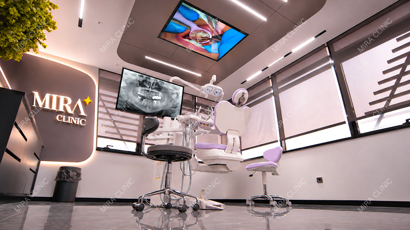
Mira Clinic has brought diagnostic technologies, clinical equipment, and its own dental laboratory together within one facility. This gives dentists and technicians the means to work on different parts of the same treatment plan while staying in direct contact.
To understand why that matters, it helps to follow the restoration’s journey.
Where Precision Begins: Understanding Each Patient’s Dental Anatomy
Before a crown can be designed, the dentist needs to understand the tooth it will restore and the teeth around it.
What shape are they? How do they meet when the patient bites? What space is available for the restoration?
Dental implant treatment brings further questions about the jawbone and surrounding anatomical structures.
Different technologies help answer different parts of this assessment.
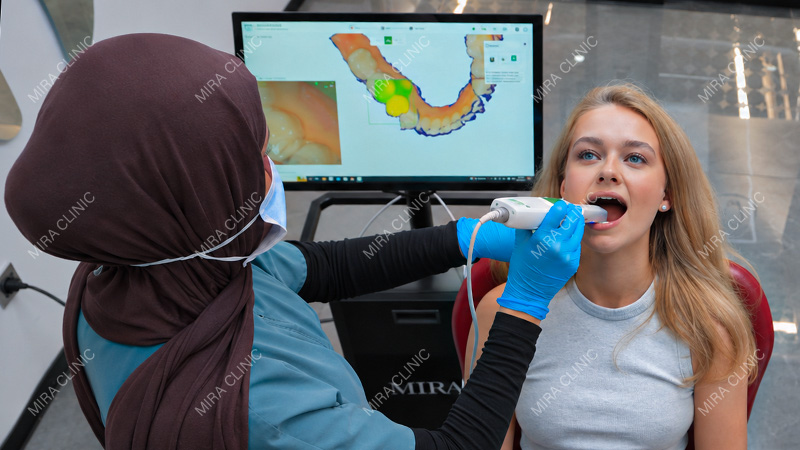
At Mira Clinic, intraoral scanners capture three-dimensional digital images of the teeth and surrounding tissues. These records allow the team to examine their shape and position and provide a starting point for restoration design.
In suitable cases, scanning also offers an alternative to conventional impressions.
But a surface scan cannot answer every clinical question.
The clinic’s diagnostic capabilities also include 2D digital radiography and 3D cone-beam computed tomography (CBCT).
For implant planning, CBCT provides three-dimensional views of bone structures and their relationship with important anatomical features. Dentists use this information to assess potential implant sites and develop a plan suited to the individual case.
A scan of the teeth and an image of the jawbone therefore serve different purposes. Choosing the appropriate records helps the dentist build the clinical picture before work moves into the laboratory.
When Implant Restorations Require Another Level of Measurement
Once implants have been placed, another measurement becomes important: their positions in relation to one another.
For a restoration supported by several implants, that information becomes part of the foundation for its design.
Mira Clinic uses dental photogrammetry to capture the three-dimensional spatial relationships between implant components in suitable cases.
The distinction from CBCT is important. CBCT helps clinicians assess anatomy and plan implant treatment. Photogrammetry records implant positions for the restorative phase.
Together, these technologies illustrate why precision involves more than taking a single detailed image. The team needs the right information at the right stage.
Beyond the Scan: Technology at the Treatment Chair
Why This Matters for International Dental Patients
Sometimes, the next step after assessment is treatment rather than design.
A tooth receiving a crown may first need root canal treatment. An implant-supported restoration follows a different sequence of clinical procedures.
The equipment used at the treatment chair reflects those differences.
During root canal treatment, the dentist works within the tooth’s narrow internal anatomy, where small details can be difficult to see.
Mira Clinic uses dental operating microscopes to magnify and illuminate this area. These can help clinicians locate hard-to-find canals, examine internal tooth structures, and manage selected complications, including separated endodontic instruments.
Magnification loupes also provide a closer view during procedures requiring detailed work.
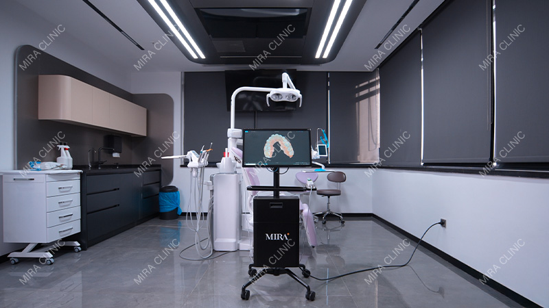
Other equipment addresses different needs. Dental laser systems are used for selected soft-tissue procedures and treatments for tooth sensitivity when clinically appropriate. Dedicated implant motors support implant procedures, while digitally equipped dental chairs with sensor-based functions form part of the clinic’s treatment facilities.
Each patient’s plan determines which of these tools is relevant.
Once the necessary clinical work is complete and the records are ready, the focus moves from treating the tooth or implant site to designing the restoration.
From Clinical Records to a Digital Restoration
A scan records what is there. Designing a restoration means deciding what needs to be created.
At Mira Clinic’s in-house laboratory, technicians use computer-aided design systems, including exocad and 3Shape, to develop crowns, veneers, bridges, and suitable implant-supported restorations.
On screen, a crown begins to acquire its contours and proportions. The technician can examine how it relates to neighboring teeth and work with the requirements provided by the dentist.
The laboratory also has extraoral scanning capabilities. When a case involves a physical dental model, that model can be digitized and brought into the design process.
Whether the starting point is an intraoral scan or a digitized model, the aim is to give the technician a detailed reference for the work ahead.
Before production begins, the design can be reviewed and refined. Three-dimensional models and restorative mockups may also be produced to help evaluate the proposed shape and proportions.
This creates an opportunity to examine the planned restoration before committing to final production.
Because the laboratory is on-site, questions can be discussed directly with the dentist. A detail about a crown’s contours or a veneer’s proportions can be considered while the design is still being developed.
Once approved for production, the design leaves the screen and becomes instructions for the manufacturing equipment.
From Digital Design to In-House Manufacturing
In the laboratory, digital information becomes something the technician can hold.
Mira Clinic uses wet and dry five-axis milling machines, alongside 3D printing systems, to produce restorations and supporting laboratory components.
For suitable crowns, bridges, and other prosthetic components, computer-aided manufacturing, or CAM, translates the approved design into instructions for milling equipment.
The machine then shapes a compatible material blank through computer-controlled cutting movements.
Five-axis milling allows tools to approach the material from different directions, supporting the production of the complex surfaces found in dental restorations.
Wet and dry milling methods accommodate different restorative materials according to the equipment’s capabilities and the material’s processing requirements.
The production method depends on what the laboratory needs to make. A final restoration, a reference model, and a prototype serve different purposes and may call for different manufacturing approaches.
How 3D Printing Complements Milling
Milling begins with a blank and removes material. 3D printing builds an object layer by layer.
At Mira Clinic, printing supports the production of dental models, restorative mockups, prototypes, and other suitable laboratory components.
A digital model can become a physical reference for inspection. A mockup can help the team assess proposed proportions before continuing with production.
These objects may not be the restoration the patient ultimately receives, but they can support the decisions involved in creating it.
Having both milling and printing available gives the laboratory different ways to turn a digital plan into physical work.
For many ceramic restorations, however, achieving the planned shape is only part of the manufacturing process.
Where Material Science Meets Dental Craftsmanship
A freshly milled zirconia restoration may already resemble a tooth. Depending on the material used, it may still need a carefully controlled heat treatment before it develops its final properties.
Mira Clinic’s laboratory works with zirconia, porcelain, and lithium-disilicate ceramic systems such as E.max.
When partially sintered zirconia is used, the milled restoration undergoes high-temperature sintering in a specialized furnace.
During this process, it shrinks.
That change must be accounted for in the digital design and manufacturing process. The shape produced by the milling machine has to allow for what will happen inside the furnace.
It is a useful example of how one stage depends on another: the technician cannot consider the design, the material, and the processing method separately.
The laboratory also uses specialized equipment for ceramic pressing, firing, and glazing. Lithium-disilicate materials, including appropriate E.max systems, provide options for veneers, crowns, inlays, and onlays.
Once the necessary processing is complete, attention turns to the details that make a restoration look natural.
Color is only one of them.
Translucency affects how light passes through the material. Surface texture influences how it reflects light. Shape and proportions determine how the restoration sits visually alongside the other teeth.
Some of these characteristics are developed during digital design. Others involve careful manual refinement and inspection by the technician.
A veneer needs to be considered alongside adjacent teeth. A crown’s appearance must also work with its functional requirements.
This is where the value of skilled finishing becomes visible. The equipment makes precise production possible; the technician evaluates and refines the details of the individual restoration.
The next stage brings that work back to the dental chair.
The Final Fitting: Bringing the Workflow Together
A restoration may look complete in the laboratory, but its final evaluation takes place during the clinical fitting.
The dentist now assesses how the finished work meets the requirements established during planning.
Three considerations come together:
- Fit: Does the restoration fit accurately?
- Function: Does it work properly with the patient’s bite and surrounding teeth?
- Appearance: Do its shape, proportions, and shade suit the patient’s smile?
A natural appearance cannot take the place of an appropriate fit or function. All three need attention before final placement.
A crown’s contours may need adjustment. A veneer may require further aesthetic refinement. An implant-supported restoration may need additional technical checks.
At Mira Clinic, the dentist can review these requirements directly with the technicians in the same building.
The clinical observations help identify what needs attention, while the design and manufacturing records help the laboratory determine how to address it.
The fitting is therefore more than the moment when the patient sees the finished restoration. It is an opportunity to evaluate the work in practice and make any necessary refinements before proceeding.
Why This Matters for International Dental Patients
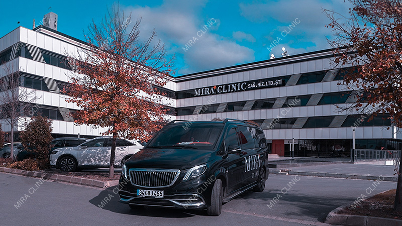
For someone traveling to Istanbul for treatment, these stages also need to fit into a practical schedule.
The patient needs to understand when to arrive, which appointments are anticipated, and whether treatment can be completed during one trip or will require separate visits.
Some laboratory work happens between appointments. Design, manufacturing, ceramic processing, finishing, and fitting each have a place in that sequence.
Mira Clinic’s in-house laboratory helps the team plan production alongside clinical appointments, with fewer transfers of restorations between separate locations.
That proximity supports coordination, but it does not remove the time required for appropriate care.
Treatment schedules still depend on the patient’s clinical condition, healing requirements, selected materials, manufacturing procedures, and quality checks. Findings during treatment or fitting may also affect the plan.
Knowing what happens between appointments gives international patients a clearer picture of their treatment: work on their restoration continues even when they are not in the treatment chair.
More Than a Collection of Advanced Machines
By the time a patient reaches the final fitting, dentists and laboratory technicians have each contributed their expertise to the restoration.
The dentist has assessed the clinical situation. The technician has developed and refined the design. Manufacturing and ceramic-processing equipment have helped turn it into a physical restoration.
The quality of the finished restoration depends on the care taken throughout that process.
At Mira Clinic, having these capabilities within one facility allows the team to follow the work from the first records through production and clinical evaluation.
For the patient, this means that the same treatment plan guides both the dentist’s clinical work and the laboratory’s decisions about how the restoration is designed, made, and finished.
Mira Clinic's digital dentistry approach is not simply the technology inside its treatment rooms or the machines inside its laboratory. It is the connection between them that brings clinical knowledge, digital precision, in-house manufacturing, and technical craftsmanship together from the first assessment to the final fit.


